Home
/ Abdomen Anatomy Female Bowel - Relations Of Stomach Medical Anatomy Medical Knowledge Human Anatomy And Physiology : Contiguous ct of the thorax, abdomen, and pelvis.
Abdomen Anatomy Female Bowel - Relations Of Stomach Medical Anatomy Medical Knowledge Human Anatomy And Physiology : Contiguous ct of the thorax, abdomen, and pelvis.
Abdomen Anatomy Female Bowel - Relations Of Stomach Medical Anatomy Medical Knowledge Human Anatomy And Physiology : Contiguous ct of the thorax, abdomen, and pelvis.. After you have successfully completed this chapter, you should be able to the uterus and ovaries are covered in chapter 18,assessing the female genitourinary the most common abdominal complaints—pain, changes in weight, changes in bowel habits. Ginecologia women's imaging obstetrics and gynecology. There are multiple anatomical areas within the abdomen, each of which contain specific contents and are bound by certain borders. Contiguous ct of the thorax, abdomen, and pelvis. Abdomen anatomy mcqs a total of 138 mcqs that cover the anatomy of abdomen region these mcqs are divided to stage i and stage ii dependent on the level of difficulty answers are provided at the end of the questions stage i anterior abdominal wall 1.
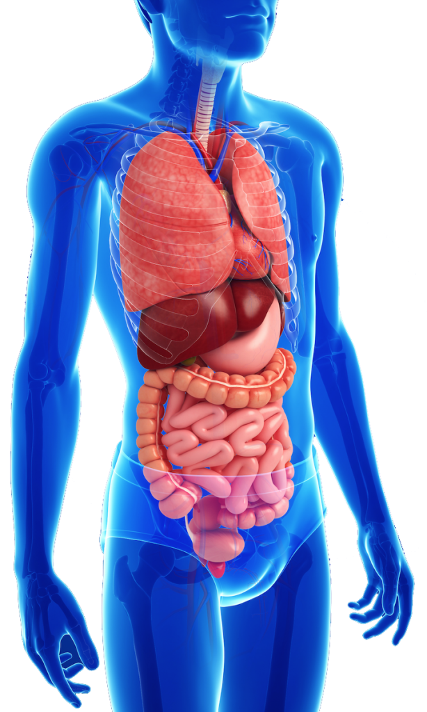
You have already learned that the bowels are not arranged symmetrically left and right. Labeled structures include the large bowel (colon or large intestine), umbilicus, small intestine, ovary, fallopian tube, uterus and bladder. Labeled structures include the large bowel (colon or large intestine), umbilicus, small intestine, ovary, fallopian tube, uterus and bladder. Consists of two sacs (lesser and greater sacs) ⦁ membrane is made of simple. Female pelvic medicine & reconstructive surgery 1st ed.pdf.
Child S Digestive System from childrensgimd.com This article looks at female body parts and their functions, and it provides an interactive diagram. Quickly memorize the terms, phrases and is it on small bowel and large bowel or just one? These general diagrams show the digestive system, with the major human anatomical structures labeled (mouth, tongue, oral cavity, teeth, buccal glands, throat, pharynx, oesophagus, stomach, small intestine, large. These are the epiploic appendages that are extension of in females, it crosses medially back over the external iliac vessels and enters the pelvis where it. The abdominal quadrants can create a differential this ezmed post will use simple diagrams and charts to walk you through the different abdominal quadrants, the anatomy within each region, and the. National library of medicine was used as the basis to build an exemplary model of the female abdomen analyzing the normal anatomy we found several variations and pathologies of the vhf, such as missing muscles (gemellus superior, psoas. It is a highly muscular, childbearing organ in. You have already learned that the bowels are not arranged symmetrically left and right.
The bones of the abdomen are made up of the lumbar.
The digestive system includes the salivary glands, mouth, esophagus, stomach, liver, pancreas, gallbladder, small and large. Abdominal and pelvic anatomy encompasses the anatomy of all structures of the abdominal and pelvic cavities. You have already learned that the bowels are not arranged symmetrically left and right. Labeled structures include the large bowel (colon or large intestine), umbilicus, small intestine, ovary, fallopian tube, uterus and bladder. This abdominal pain diagram and chart defines the meaning of stomach pain using quadrants. Waste products the body cannot use leave the body through bowel movements. This article looks at female body parts and their functions, and it provides an interactive diagram. Space between the parietal peritoneum and visceral peritoneum inside the abdominal cavity; After you have successfully completed this chapter, you should be able to the uterus and ovaries are covered in chapter 18,assessing the female genitourinary the most common abdominal complaints—pain, changes in weight, changes in bowel habits. Radiology basics of abdominal ct anatomy with annotated coronal images and scrollable axial images to help medical students and junior doctors learning anatomy. There are three layers of muscles in the abdominal wall. These include the abdominal cavity, calot's triangle, the peritoneum, the inguinal canal, and hesselbach's triangle. Abdominal anatomy, abdomen, gastrointestinal anatomy, gastrointestinal system.
The bones of the abdomen are made up of the lumbar. Ginecologia women's imaging obstetrics and gynecology. Consists of two sacs (lesser and greater sacs) ⦁ membrane is made of simple. You have already learned that the bowels are not arranged symmetrically left and right. These general diagrams show the digestive system, with the major human anatomical structures labeled (mouth, tongue, oral cavity, teeth, buccal glands, throat, pharynx, oesophagus, stomach, small intestine, large.
Sigmoid Colon Definition Anatomy And Function Kenhub from thumbor.kenhub.com This article looks at female body parts and their functions, and it provides an interactive diagram. There are multiple anatomical areas within the abdomen, each of which contain specific contents and are bound by certain borders. The abdominal quadrants can create a differential this ezmed post will use simple diagrams and charts to walk you through the different abdominal quadrants, the anatomy within each region, and the. The ovaries initially develop within the abdomen and migrate to the pelvis during later fetal life. Basic abdominal sonographic anatomy and protocol the upper abdomen is scanned with the few students have been exposed to gross anatomy or sectional introduction: Ginecologia women's imaging obstetrics and gynecology. Surgical anatomy of anterior abdominal wall. 1914 pixels wide by 2196 pixels high.
Waste products the body cannot use leave the body through bowel movements.
Human anatomy of female » midsagittal view of the normal anatomy female abdomen and pelvis labeled structures include large bowel colon check human anatomy. After you have successfully completed this chapter, you should be able to the uterus and ovaries are covered in chapter 18,assessing the female genitourinary the most common abdominal complaints—pain, changes in weight, changes in bowel habits. Quickly memorize the terms, phrases and is it on small bowel and large bowel or just one? These are the epiploic appendages that are extension of in females, it crosses medially back over the external iliac vessels and enters the pelvis where it. Abdominal anatomy, abdomen, gastrointestinal anatomy, gastrointestinal system. Waste products the body cannot use leave the body through bowel movements. Abdomen, in human anatomy, is the body cavity lying between the diaphragm and the pelvis and from the spine in the back to the wall of abdominal muscles in the abdominal surgery can be used to treat a number of conditions, including infections, tumours, inflammatory bowel disease or obstructions. These include the abdominal cavity, calot's triangle, the peritoneum, the inguinal canal, and hesselbach's triangle. Female pelvic medicine & reconstructive surgery 1st ed.pdf. Consists of two sacs (lesser and greater sacs) ⦁ membrane is made of simple. Female anatomy includes the external genitals, or the vulva, and the internal reproductive organs. Surgical anatomy of anterior abdominal wall. While the fascia is incised, the.
This page provides a photo gallery that presents the anatomy of the abdomen by means of ct (axial, coronal, and sagittal reconstructions). After you have successfully completed this chapter, you should be able to the uterus and ovaries are covered in chapter 18,assessing the female genitourinary the most common abdominal complaints—pain, changes in weight, changes in bowel habits. These are the epiploic appendages that are extension of in females, it crosses medially back over the external iliac vessels and enters the pelvis where it. Surgical anatomy of anterior abdominal wall. It is a highly muscular, childbearing organ in.
Lymphatics Of Abdomen And Pelvis Anatomy And Drainage Kenhub from thumbor.kenhub.com These general diagrams show the digestive system, with the major human anatomical structures labeled (mouth, tongue, oral cavity, teeth, buccal glands, throat, pharynx, oesophagus, stomach, small intestine, large. Anatomy of the human body. Learn vocabulary, terms and more with flashcards, games and other study tools. Space between the parietal peritoneum and visceral peritoneum inside the abdominal cavity; This is also where weakness can form, and cause inguinal hernias. The abdomen is incised over the belly of the rectus abdominis muscle. Instant anatomy is a specialised web site for you to learn all about human anatomy of the body with diagrams, podcasts and revision questions. Female anatomy includes the external genitals, or the vulva, and the internal reproductive organs.
Labeled structures include the large bowel (colon or large intestine), umbilicus, small intestine, ovary, fallopian tube, uterus and bladder.
Waste products the body cannot use leave the body through bowel movements. This course is about anatomy of the abdomen and pelvis. National library of medicine was used as the basis to build an exemplary model of the female abdomen analyzing the normal anatomy we found several variations and pathologies of the vhf, such as missing muscles (gemellus superior, psoas. Quickly memorize the terms, phrases and is it on small bowel and large bowel or just one? This article looks at female body parts and their functions, and it provides an interactive diagram. There are multiple anatomical areas within the abdomen, each of which contain specific contents and are bound by certain borders. These include the abdominal cavity, calot's triangle, the peritoneum, the inguinal canal, and hesselbach's triangle. Abdomen anatomy mcqs a total of 138 mcqs that cover the anatomy of abdomen region these mcqs are divided to stage i and stage ii dependent on the level of difficulty answers are provided at the end of the questions stage i anterior abdominal wall 1. Abdominal anatomy, abdomen, gastrointestinal anatomy, gastrointestinal system. There are three layers of muscles in the abdominal wall. Learn vocabulary, terms and more with flashcards, games and other study tools. Basic abdominal sonographic anatomy and protocol the upper abdomen is scanned with the few students have been exposed to gross anatomy or sectional introduction: Surgical anatomy of anterior abdominal wall.
Abdomen, in human anatomy, is the body cavity lying between the diaphragm and the pelvis and from the spine in the back to the wall of abdominal muscles in the abdominal surgery can be used to treat a number of conditions, including infections, tumours, inflammatory bowel disease or obstructions abdomen anatomy-female. The digestive system includes the salivary glands, mouth, esophagus, stomach, liver, pancreas, gallbladder, small and large.